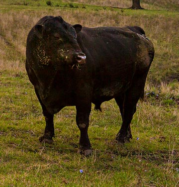
Good reproductive performance of a bull (Figure 1) is necessary to obtain a high percent calf crop when natural service is used for breeding. A bull must be fertile, capable and willing to mate a large number of cows during a short breeding season for optimum production. A basic knowledge of the reproductive tract is beneficial for improved management. An understanding of the bull’s reproductive system will also help the producer better understand breeding soundness examinations, reproductive problems and breeding impairments.
Anatomy
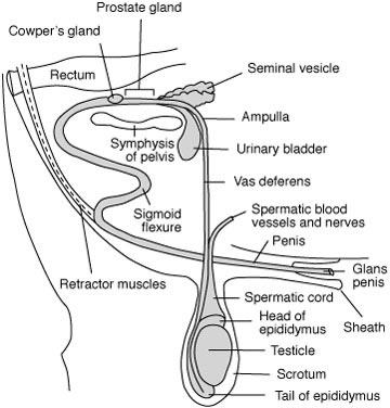
The reproductive tract of the bull consists of the testicles, secondary sex organs, and three accessory sex glands. These organs work in concert for formation, maturation and transport of spermatozoa, which are eventually deposited in the female reproductive tract. The secondary sex organs are the epididymis, vas deferens and penis. The three accessory sex glands include the seminal vesicles, prostate and bulbourethral gland (Cowper’s gland). This basic anatomy is illustrated in Figure 2.
Testicle
The testicle is located outside the body cavity in the scrotum and has two vital functions: producing the spermatozoa, and producing the male hormone, testosterone. Location of the testicles exterior to the body cavity is essential for normal sperm formation, which occurs only at 4 degrees to 5 degrees below body temperature. The scrotum provides physical protection to the testicle and helps regulate the temperature for optimum spermatozoa development. This regulation is done by coordination of three structures: a temperature-sensitive layer of muscle (tunica dartos) located in the walls of the scrotum, which relaxes when hot and contracts when cold; the external cremaster muscle within the spermatic cord, which controls the proximity of the testicle to the body by lengthening or shortening depending on environmental temperature; and a counter-current temperature exchange regulated by a blood flow process known as the pampiniform plexus, which is a coil of testicular veins that provide an effective mechanism for cooling arterial blood entering the testicle and transferring its heat to the venous blood leaving the testicle.
One or both testicles occasionally fail to descend into the scrotum during embryological development and are retained in the body cavity. This condition is known as chryptorchidism. Hormone production by chryptorchid males is near normal and the male develops and behaves like a normal male, but will generally be subfertile. This condition is genetically inherited, therefore such males should not be used for breeding.
The testicle contains many long, tiny, coiled tubes known as seminiferous tubules, within which the sperm are formed and begin to mature. Scattered throughout the loose connective tissue surrounding the seminiferous tubules are many highly specialized cells, the interstitial cells of Leydig, that produce testosterone. There are hundreds of individual seminiferous tubules in the body of the testicle which unite with one another to form a few dozen tubules that exit from the testicle and pass into the epididymis.
Epididymis
The epididymis is a compact, flat, elongated structure closely attached to one side of the testicle. It is divided into three regions, the head, body and tail. The many tubules entered the head of the epididymis from the testicle unite to form a single tubule some 130 to 160 feet in length. This tubule is convoluted and packed into the 6- to 8-inch epididymis. Four major functions occur in the epididymis, including the transport of the developing sperm cells from the testicle to the vas deferens; the concentration of the sperm by absorption of surplus fluids; the maturation of the developing spermatozoa; and the storage of viable sperm cells in the epididymis tail. If sexual activity is slowed, resorption of sperm cells from the epididymis tail occurs.
The epididymis serves as an outlet for all the sperm produced in the testicle and any blockage of this tube will cause sterility. Temporary blockage due to swelling following an injury or infection (epididymitis) will result in short-term infertility. If the swelling or infection results in formation of scar tissue in the tubule, it may permanently block the passage of sperm. If blockage occurs in both epididymides, the bull will no longer be useful as a breeder. Surgical removal of the tail of the epididymis (epididectomy) is frequently used as a means of sterilization for teaser (Gomer) bulls for estrus detection. Epididectomized bulls will still service cows in the usual manner, but will not deposit sperm in the female reproductive tract.
Vas deferens
The vas deferens, also known as ductus deferens, emerges from the tail of the epididymis as a straight tubule and passes as part of the spermatic cord through the inguinal ring into the body cavity. Spermatozoa are transported further along the reproductive tract to the pelvic region through the vas deferens by contraction of the smooth muscle tissue surrounding this tubule during ejaculation. Bulls may also be sterilized by a vasectomy in which a section of the vas deferens is removed so that sperm cannot pass to the outside of the body.
Urethra
The two vas deferens eventually unite into a single tube, the urethra, which is the channel passing through the penis. The urethra in the male serves as a common passageway for semen from the reproductive tract and urine from the urinary tract.
Accessory glands
Two of the accessory glands are found in the general region where the vas deferens unite to become the urethra. Secretions from these glands make up most of the liquid portion of the semen. In addition, the secretions activate the sperm to become motile. The seminal vesicles consist of two lobes about 4 to 5 inches long, each connected to the urethra by a duct. The prostate gland is located at the neck of the urinary bladder where it empties into the urethra. The prostate is relatively small in the bull, as compared to other species, and does not produce a very large volume secretion.
The third accessory gland, the Cowper’s glands, are small, firm glands located on either side of the urethra. The clear secretion that often drips from the penis during sexual excitement prior to service is largely produced by these glands and serves to flush and cleanse the urethra of any urine residue that may be harmful to spermatozoa.
One of the accessory glands may occasionally become infected, resulting in semen samples that are yellow and cloudy and which contain puss cells. It is not uncommon in bulls for the seminal vesicles to be so affected (seminal vesiculitis). Antibiotic treatment is sometimes necessary, but time will generally correct the problem.
Penis
The sigmoid flexure is an anatomical structure that provides a means by which the penis is held inside the sheath except during time of service. Strong retractor muscles hold the penis in the “S” shaped configuration. Occasionally these muscles are too weak to function properly and a portion of the penis and sheath lining protrude at all times. This exposes the male to the danger of injury and this characteristic should be avoided when selecting a herd bull.
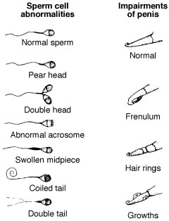
The penis is the organ of insemination. Spongy-type material within the penis is filled with blood during sexual arousal, resulting in erection of the organ. The end of the penis is the glans penis and is richly supplied nerves, which are stimulated during copulation to induce ejaculation. Impairments of the glans penis may exist (Figure 3) and should be detected during a fertility exam.
Regulation of male hormones
The normal functions of male reproduction are largely controlled by hormones that are secreted from the endocrine glands. The testicle functions as an endocrine gland because of its production of the male hormone, testosterone, by the intersticial cells. Testosterone has several major functions:
- It is largely responsible for development and maintenance of the male reproductive tract
- It causes the development and maintenance of the secondary sex characteristics associated with masculinity, such as the crest and heavily muscled shoulders of a bull
- It is a major factor in the normal sex drive and behavior of the male
- It increases muscular and skeletal growth
- It is essential for normal sperm formation.
The testicle is, in turn, under the influence of hormones produced by other glands in the body. The same gonadotropic hormones that regulate ovarian functions in the cow also regulate testicular functions in the bull. Luteinizing hormone (LH) and follicle stimulating hormone (FSH) appear to be misnamed as pertaining to male reproduction, yet carry out several important male functions.
LH and FSH are released from the pituitary gland and cause the testicle to secrete testosterone, which then acts on the germ cell lining of the seminiferous tubules to stimulate formation of primordial sperm cells. The maturation of spermatids into fully developed sperm cells requires the presence of FSH. Normal functioning of the male accessory glands requires testosterone.
Not only is hormone production of the testicle regulated by hormones released by the anterior pituitary, but the reverse is also true. The level of testosterone in the blood regulates the secretion of gonadotropic hormones from the anterior pituitary via a feedback system. A proper balance of all hormones is vital to successful reproductive functions.
Breeding soundness exams
An examination of bulls for breeding soundness before the breeding season can detect the majority of bulls which have obvious potential fertility problems. This examination should be performed by an experienced, trained person, usually a veterinarian. It should be pointed out here that a breeding soundness examination is simply a screening procedure to eliminate bulls that have a high possibility of being non-fertile. A “fertility test” is a misnomer since techniques presently available do not allow for accurate prediction of fertility. Results from an actual breeding season remain the only test of a bull’s breeding ability. For more detailed information on bull breeding soundness exams, refer to MU Extension publication G2011, Determining Reproductive Fertility in Herd Bulls. A breeding soundness examination includes an evaluation of the following components:
Physical examination
The penis should be examined during electro-ejaculation or natural mating, in an erected, extended state. Figure 2 illustrates possible abnormalities of the penis. Potential problems of the penis may include hair rings, which restrict circulation, a persistent frenulum or adhesion, lacerations, growths, scar tissue, deviations, or a urethral fistula. Next the scrotum and testes should be palpated. The scrotum should be pendulous but well supported. The testes should be firm and uniform in size and shape. The internal sex organs should be palpated rectally to ensure proper development and size. Good vision and sound feet and legs should also be considered when evaluating a bull’s physical abilities to breed.
Scrotal circumference
Scrotal circumference gives an indication of a bull’s ability to produce sperm and is related to younger age at puberty. Breeds differ somewhat as to scrotal circumference, but 32 centimeters is generally accepted as the minimum size for yearling bulls to be sound breeds.
Semen evaluation
Four criteria are used to evaluate semen, i.e., volume, concentration, motility and morphology. Proper training is required to accurately evaluate semen. A scoring system for predicting potential breeding soundness of bulls as prepared by the Society for Theriogenology, which incorporates scrotal circumference, semen motility, and semen morphology.
Breeding soundness examinations presently do not include an evaluation of a bull’s sexual drive or libido. Libido testing is time consuming and requires use of females in estrus and therefore is difficult to conduct on a large scale. Procedures are being investigated to better evaluate the willingness and desire of bulls to mate.
Portions of this guide were adapted from Nebguide G80-536, by G.H. Deutscher, Extension Beef Specialist, University of Nebraska.