Pregnancy examination provides valuable information, as it can enable cow-calf operations to make management decisions that both increase profitability and improve long-term reproductive efficiency. Several methods of pregnancy examination may be considered based on the goals of individual operations and the availability of the method.
Value of pregnancy examination
When performed at an appropriate time, pregnancy examination (Figure 1) can provide valuable information to make management decisions in beef herds.
Cost reduction
Non-pregnant or “open” females that fail to conceive within a defined breeding season are often a significant source of profit loss. While selling otherwise sound animals can be a difficult decision for many producers, carrying nonproductive animals until the next breeding season can cost hundreds of dollars in feed and other management costs. Culling these cows prevents unnecessary losses and directly increases profit for the current year.
Managing reproductive efficiency
Pregnancy examination can also be used as a tool to improve reproductive efficiency through identification of late-conceiving females. Cows that conceive later in the breeding season will calve later and therefore have a shorter period of time to resume normal estrous cycles before the next breeding season. Strategically marketing late-conceiving animals can improve the reproductive performance of the herd in the next breeding season, shorten the calving season, and result in a more uniform and profitable calf crop.
Additionally, identifying heifers that conceive early in the breeding season aids in selecting replacement animals that match the reproductive schedule of the herd. Heifers that conceive early in their first breeding season remain in the herd for a greater number of years and also wean older, heavier calves each year. In many cases, marketing later-conceiving heifers following early pregnancy examination is more profitable than retaining these heifers as replacements.
Pregnancy examination as a diagnostic tool
Pregnancy rate may be used as indicator of reproductive performance over time or as a tool to diagnose sources of reduced reproductive performance. Comparing pregnancy status over multiple years allows for evaluation of changes in reproductive management. Poor pregnancy rates warrant an investigation of potential causes for unsatisfactory performance. Accurate information about pregnancy attainment informs management decisions regarding nutrition, animal health, estrus synchronization programs, or bull selection for future years.
Methods of pregnancy examination
Rectal palpation
Rectal palpation is a simple, yet effective method of detecting pregnancy when performed by a trained and experienced individual. In this method, palpation of the reproductive tract through the rectal wall allows the detection of a number of changes that accompany pregnancy. Signs such as asymmetrical uterine horns, fluid in the uterus, structures on the developing placenta, or size of the fetus indicate both the presence and stage of a pregnancy. Accurate assessment of these signs requires considerable skill, so palpation should only be performed by experienced professionals.
Pregnancy can be identified as early as 35 days of gestation by skilled palpators, but prediction of fetal age is most accurate between 45 and 120 days. The size of placentomes (attachment points between the placenta and uterus) provides another indication of fetal age between 90 and 150 days, although fetal age estimates made based on placentome size are less precise. Although it is the simplest form of examination, rectal palpation is limited in that it cannot assess qualities such as fetal viability or fetal sex.
Ultrasound
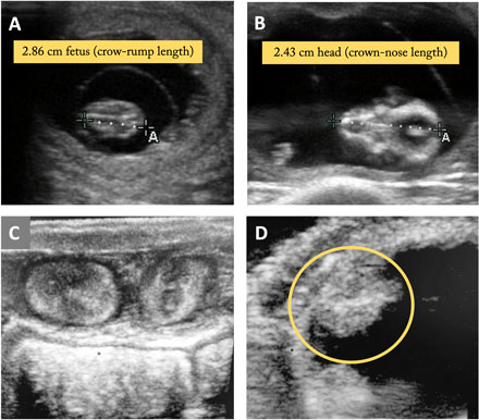
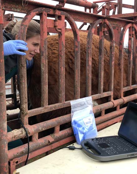
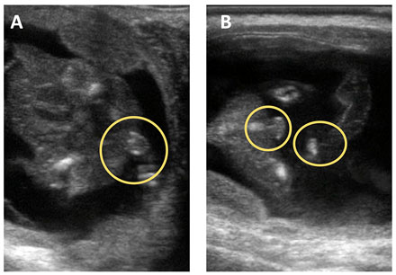
Ultrasound provides a more conclusive and comprehensive assessment of pregnancy. Images of the uterus and surrounding structures (Figure 2) are obtained through manipulation of an ultrasound probe inserted in the rectum. This method allows for earlier detection at around 28 days of gestation. Ultrasound requires less experience and does not depend as heavily on technician skill, reducing errors in diagnosing pregnancy. Perhaps the greatest benefit is the amount of information provided, as ultrasound allows assessment of fetal viability, identification of reproductive or developmental abnormalities, determination of fetal sex (Figure 3), and a more precise determination of fetal age and expected calving date.
Biochemical tests
Advances in biochemical technology have resulted in the development of commercially available tests for pregnancy status. These tests are capable of identifying hormone patterns or proteins specific to pregnancy. Common tests include those for pregnancy-associated glycoproteins (PAGs) or progesterone concentrations in samples of blood or milk. The mechanism of each test dictates the type of sample required and the speed at which results are obtained, as many tests require shipping of samples to laboratories for analysis. Recently, increasing availability of on-farm tests that provide immediate results has made biochemical tests a more attractive option. In most cases, sampling requires minimal training, yet tests are highly accurate for detection as early as 25 days. Unfortunately, no additional information is provided about the pregnancy or when conception occurred, unless timing of testing is done strategically. Therefore, biochemical tests may not be the most effective option for all commercial operations.
Table 1. Day of gestation and observations during a pregnancy examination.
| Day of gestation | Observation | Characteristics |
|---|---|---|
| 35 | Vesicle approximately 2 cm Fetus approximately 1 cm |
Uterus slightly enlarged and filled with fluid |
| 45 | Vesicle approximately 4 cm Fetus approximately 3 cm |
One uterine horn enlarged |
| 60 | Fetal head approximately 2.5 cm long (crown to nose) Fetus approximately the size of a mouse |
Fetal sex determination possible via ultrasound based on location of genital tubercle |
| 75 | Fetal head approximately 3.5 cm long (crown to nose) | Scrotum and teats visible via ultrasound; placentomes approximately pea-sized |
| 90 | Fetal head approximately 5 cm long (crown to nose) Fetus approximately the size of a rat |
Uterus often over the pelvis and becoming more difficult to reach; placentomes dime-sized |
| 120 | Fetus approximately the size of a small cat | Uterus generally deep within body cavity and difficult to reach; placentomes quarter-sized |
| 150 | Fetus approximately the size of a large cat | Fremitus may be felt in the uterine artery |
| 180 | Fetus approximately the size of a small dog | Growing calf is now more easily reached and movement may be felt |
Timing
Pregnancy examinations need to be performed at the appropriate time to capture important information (Figure 4). Earlier detection allows cost-saving culling decisions to be made sooner. However, performing pregnancy examination earlier also has limitations. Pregnancy examinations should be scheduled to assess desired characteristics at the appropriate point of gestation relative to artificial insemination or bull turn out.
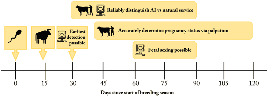
Ultrasounds must be conducted somewhat early in gestation to determine some characteristics, as the size and depth of the fetus will be too great to allow complete visualization later in pregnancy. Fetal sex may be determined reliably from about 60 to 90 days of gestation by pinpointing the location of the genital tubercle or identifying secondary reproductive structures, such as the scrotum of bull calves. Although fetal sex can be determined after day 90 as well, it is more difficult.
Knowing the possible range in days of gestation is helpful for identifying pregnancy rates to AI and determining fetal age via palpation or ultrasound. Additional timing considerations may be necessary for differentiation of AI and natural service sired calves. If this is a priority, consider waiting two weeks before bull turn out following AI to ensure a clear distinction can be made Ensure pregnancy examination is performed prior to 90 days after AI as natural variation in fetal size and depth of the pregnancy within the animal can make it difficult to accurately distinguish AI pregnancies from pregnancies that result from natural service after this point.
Summary
Pregnancy examination can provide valuable information to make management decisions or to identify challenges and opportunities for improvement. Consult your veterinarian for the most advantageous timing for pregnancy examinations in your operation.