Current samples
Volutella Leaf Blight of Spurge
Riley France and Peng Tian
Japanese pachysandra, also known as Japanese spurge, is a popular ornamental plant used as groundcover in shady areas. This fragrant-blooming plant can help reduce erosion and keep areas cool while enhancing their aesthetic appeal. However, as spring rains encourage their growth, the dense planting of this Pachysandra also encourages the growth of an unsightly pathogen – Volutella leaf blight.
Symptoms: Volutella leaf blight infections start out mild and can go undetected for years. It manifests as tan to dark grayish, water-soaked lesions on both stems and leaves. As stem lesions age, they will dry and form dark, cracked cankers of dead tissue that cut off water and nutrients to the rest of the stem. Leaf lesions grow in size, developing irregular concentric rings. In particularly wet seasons or if the plant is experiencing other stresses, the severity of the infection can increase, resulting in dead patches in the stand.

Learn more about the life cycle and management of this disease:
Tomato Spotted Wilt Virus
Riley France and Peng Tian
Originally discovered in Australia in 1915 and found on U.S. soil in the 1970’s, tomato spotted wilt virus (TSWV) is a highly impactful plant disease that can result in both aesthetic damage and significant loss in crop revenue. This Tospovirus has one of the widest known host ranges, with over 1000 susceptible species in 85 families. As it is unable to move on its own, TSWV is primarily transmitted by thrips. Due to the easy spread, broad host range, and economical loss caused by TSWV, it is important to understand how to identify and manage infected crops.
Symptoms: The symptoms of TSWV may vary from different host species or different cultivars within a single host species. The plants frequently appear as stunted, wilting, bronzing with cupping of younger leaves, and chlorotic and necrotic spots with or without a bulls-eye pattern. Symptoms can also manifest on fruit, frequently as ring-shaped chlorotic blotches.

Learn more about the life cycle and management of this disease:
Orange Rust of Brambles
Peng Tian and Blaike Zehnle
Orange rust is a notable fungal disease impacting bramble species, particularly blackberries and certain raspberry cultivars. The disease, caused by fungi in the genus Arthuriomyces and Gymnoconia, is notorious for its ability to cause systemic infections that can severely diminish vegetative growth of affected plants and fruit production, leading to substantial economic losses. Therefore, a comprehensive understanding of the disease's symptoms, life cycle, and management strategies is critical for growers seeking to sustain healthy and productive bramble crops.
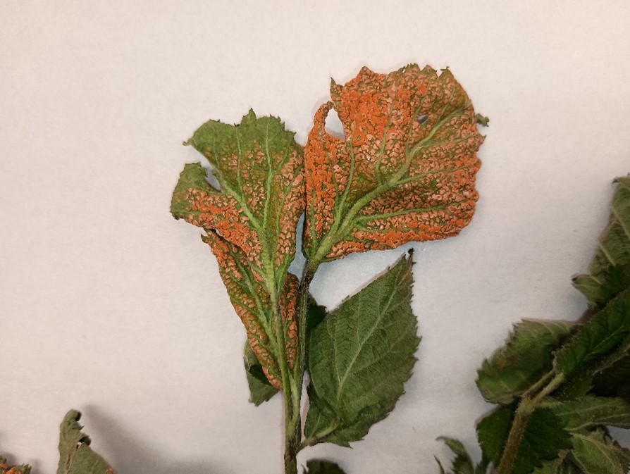
Symptoms and signs: The first symptoms of orange rust appear in early spring when new shoots and leaves emerge. Infected plants exhibit pale, yellowish-green leaves that may appear stunted or deformed. Black specks surrounded by chlorotic tissue will be present on the underside of the leaves. As the disease progresses, the characteristic orange, waxy pustules develop from the black specks (Figure 1). These pustules release bright orange, powdery spore masses that are easily spread by wind. Infected plants typically exhibit weak growth and produce fewer or no fruit as the disease systemically affects the entire plant.
Learn more about the diagnosis and management of this disease:
Rhizoctonia Bottom Rot of Lettuce
Peng Tian and Blaike Zehnle
Bottom Rot caused by Rhizoctonia solani was confirmed from a lettuce sample received by MU Plant Diagnostic Clinic. This soilborne fungus is known for causing damping-off and bottom rot on a broad range of plants when they approach maturity, both in open fields and under shelter. It is most economically damaging during harvest when growers must trim plants with rotten leaves, reducing both the quality and yield.
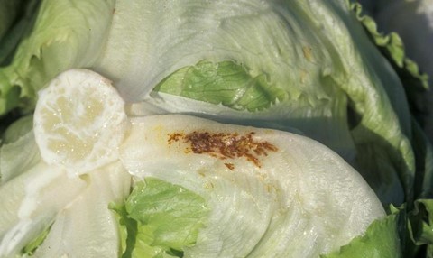
Symptoms and signs: Bottom rot symptoms typically develop first on lower leaves in contact with the soil and appear as small, rust-colored brown spots, mainly under leaf midribs (Figure 1). Symptoms generally are most pronounced at heading. Bottom rot can spread to midribs and leaf blades rapidly when conditions are favorable. Stems are relatively more resistant to bottom rot and are the last portion of the head to decay. Decaying heads are at first slimy and brown but become dark brown to black as they collapse and dry. A webbed network of white to brown mycelium often grows over lesions, and small gray, brown sclerotia later are apparent. Botrytis cinerea and bacteria may begin to colonize on the damaged tissues and accelerate the rotting.
Learn more about the diagnosis and management of this disease:
Bur Oak Blight
Peng Tian and Pierce Taylor
Across the Midwest, Bur Oak Blight (Tubakia iowensis) is a fungal pathogen infecting the Bur Oak (Quercus macrocarpa). Affected plants display chlorosis and fruiting bodies on the leaf vein and petiole in mid to late summer. The small-acorn variety of bur oak (Quercus macrocarpa var. oliviformis), is affected more severely than other varieties. The disease can reinfect trees year to year and can eventually cause death after several years of defoliation.
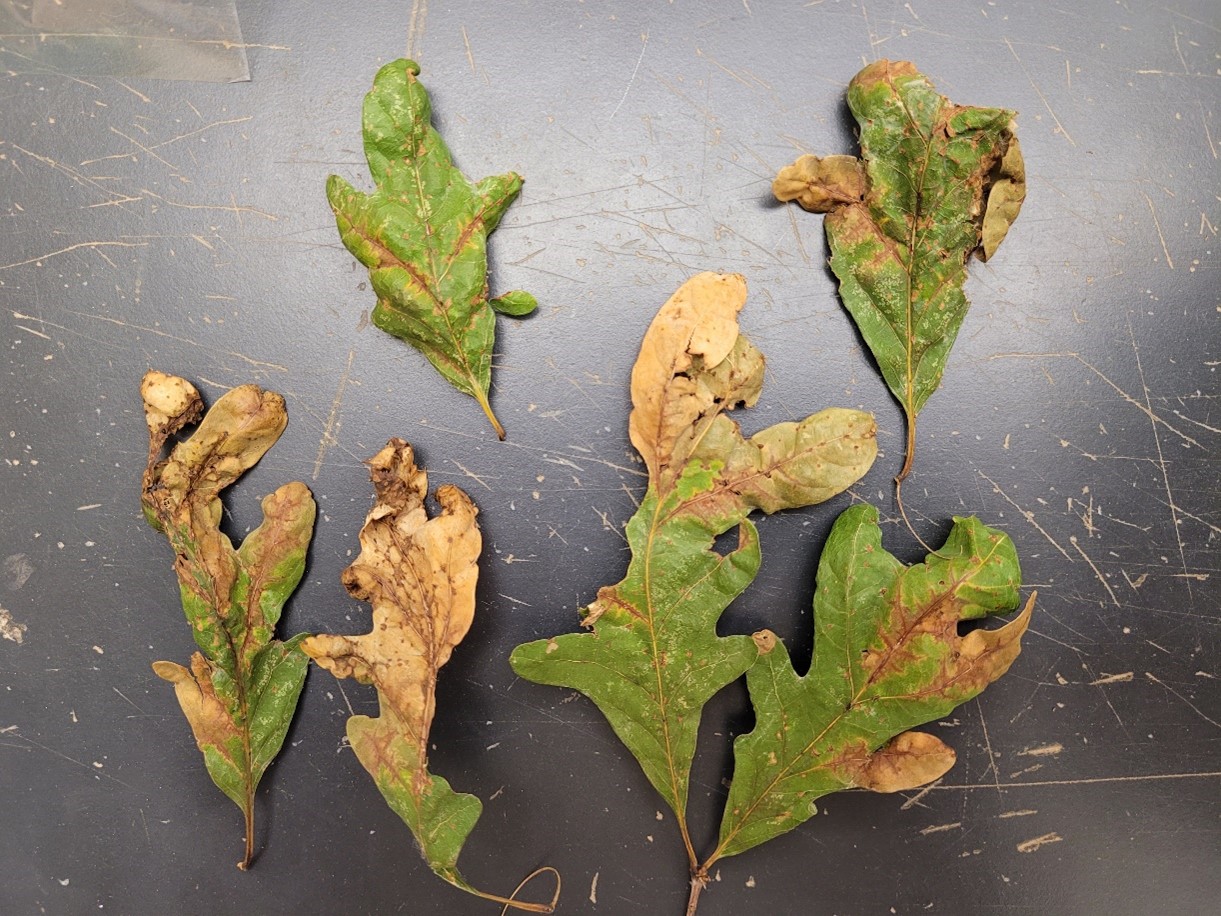
Symptoms and signs: Typical symptoms include necrosis(death) occurring around the leaf veins, as well as the tip. Eventually the fungus occurring on leaf veins result in tissue death. The presence of fruiting bodies can be seen along the veins on the underside of the leaf and around the petiole as a purple discoloration. Due to the death of foliage, Bur Oak Blight is often confused with Oak wilt or Anthracnose disease. These fruiting bodies, or pycnothyrium, presence on the petiole and the tree retaining its leaves over winter are symptoms of Bur Oak Blight which differentiate it from similar diseases. The conidia spores of Tubakia iowensis are microscopic and cannot be seen unaided, thus proper identification requires analysis with a laboratory microscope. Tubakia spp. have a pycnothyrium that composes of a shield like scutellum of radial projected hyphae. The scutellum is supported by a short columnar stalk that bears the round to ovoid conidia spores.
Learn more about the life cycle, damage and management of this disease: